

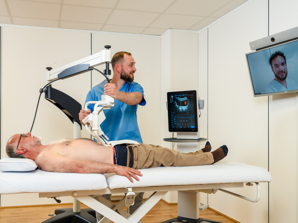
Robotic tele-ultrasound allows a remote expert to perform an ultrasound examination using a robotic arm. Meanwhile, an operator on site supports the patient and positions the device. But how does a tele-ultrasound robot actually work? How are the expert’s movements and the ultrasound images transmitted in real time? In this article, discover robotic tele-ultrasound and the technology behind it.
Robotic tele-ultrasound: technology and how it works
Robotic tele-ultrasound is a technology that comes in three main families: the robotic arm, the motorised probe and assisted remote guidance.
- Robotic arm (RA): an articulated arm holds a standard ultrasound probe and reproduces the expert’s movements remotely.
- Motorised probe (MP): the probe contains micro-motors and the remote expert controls the orientation of the transducer inside the probe.
- Remote guidance (RG): a professional on site holds the probe and is guided in real time by an expert via video conference.
Each of these three architectures meets different needs, while offering diagnostic quality comparable to conventional ultrasound (P. Arbeille et al., 2016).
AdEchoTech designs robotic tele-ultrasound systems for performing ultrasound examinations remotely. Discover the MELODY robotic tele-ultrasound solution.
The robotic arm: the first generation of robotic ultrasound
MELODY was born from the development of the robotic arm, the first tele-ultrasound technology validated in clinical practice through European projects and space applications. This initial success laid the foundations for what MELODY is today: a tangible technology deployed to address the challenges of access to care.
An articulated arm mounted on a support holds a standard probe (abdominal, cardiac or linear). A non-expert caregiver places the arm over the area to be examined and holds it in position. An expert, in another location, handles a dummy probe. Their movements (tilting, rotation or small displacements) are reproduced by the arm on the patient. The ultrasound images are sent as a video stream, often accompanied by a video conference to communicate with the patient and the caregiver.
AdEchoTech’s MELODY system illustrates this approach. This technology gives the expert full control over the orientation of the probe. It is compatible with a wide range of standard probes, which makes it easy to integrate into existing equipment. It has been validated in a variety of settings: medical deserts, emergency care, pregnancy monitoring, crisis situations and even space ultrasound aboard the ISS.
Studies show that the robotic arm makes it possible to reach a diagnosis in around 97% of cases for deep organs, with image quality close to that of conventional ultrasound.
On the other hand, the arm and its support are bulky and heavy, which makes certain movements and positions more difficult. A caregiver is needed on site to position and stabilise the arm, and sometimes to adjust the ultrasound scanner. Examinations take longer on average than with a motorised probe, especially for deep organs, because small translational movements are less precise.
Despite these limitations, the robotic arm, and in particular AdEchoTech’s MELODY solution, remains a reference solution for centres that want versatile tele-ultrasound compatible with their current probes.
Motorised probe: robotics built into the probe
The motorised probe marks the arrival of the second generation of tele-ultrasound. It was designed to be more ergonomic and easier to deploy.
The probe has small motors that tilt and rotate the transducer. A caregiver places the probe on the area to be examined and holds it in place. The remote expert uses a dummy probe and a keyboard to orient the transducer. Some solutions, such as those from Sonoscanner or certain CNES/ESA projects, use this principle.
This architecture offers excellent ergonomics. The probe is light and easy to position under the ribs or in areas that are difficult to access. Examination times are shorter for deep organs and significantly faster than with a robotic arm (P. Arbeille et al., 2016).
Expert mobility is also a strong point. All that is needed is a computer and a USB dummy probe, which can be used from a practice, from home or from a vehicle, as long as the Internet connection is secure. It is also possible to acquire 3D volumes and reanalyse them later at the expert centre.
This technology does however have a few constraints. It requires specific motorised probes, which are fewer in number and sometimes more expensive than standard probes. The head of the motorised probe may be less suitable than the very high-frequency linear probes used in remote guidance for certain very superficial and fine vessels.
The motorised probe is now considered the best compromise for deep organs and volumetric superficial organs such as the thyroid, muscles or abdomen. It is also highly reliable from a diagnostic point of view.
Assisted remote guidance: the importance of caregiver training
Remote guidance is the simplest solution technically, but it requires good training of the caregiver on site. A professional, often a general practitioner, holds the probe and performs the examination. An expert guides them live via video conference, indicating where to place the probe, how to orient it and which settings to apply.
Remote guidance is suited to superficial vessels, such as the carotid arteries or the veins of the legs. The diagnostic rate is around 98% for these indications. A non-specialist learns to obtain the necessary transverse and longitudinal views with about one week of training.
However, remote guidance alone is not suited to deep organs such as the liver, kidneys, pancreas, pelvis or foetus, nor to certain volumetric superficial organs.
Assisted remote guidance therefore has its place as a complement to robotic solutions for examinations such as superficial arterial or venous Doppler ultrasound, and in facilities that already have conventional ultrasound scanners.
Data transmission: the backbone of the system
The quality of tele-ultrasound depends on the transmission of data such as the ultrasound images, the robot commands, and the ambient sound and video.
The robot commands represent little data, but they are highly sensitive to delay. If latency is too high, remote manipulation becomes unresponsive. The ultrasound video is a real-time stream that requires good bandwidth and must preserve clinical details, whether in B-mode, colour Doppler or spectral Doppler. Ambient audio and video are useful for talking to the patient and the caregiver.
Work on robotic cardiac tele-ultrasound has shown that commercial 4G-LTE networks now allow very low latencies, compatible with responsive remote manipulation. They offer bandwidths of several Mbps, sufficient to transmit good-quality ultrasound video with modern codecs such as H.264/AVC or HEVC without diagnostic loss. The user experience is close to that of an in-hospital ultrasound with bandwidths of around 2 Mbps.
These performances pave the way for deployments in rural areas, in ambulances or in emergency situations. Fibre, 5G and satellite links complete the picture, whether for hospitals, health centres, isolated sites or space missions.
MELODY in practice: a solution already deployed in routine care
Unlike many research prototypes, MELODY is a commercial solution already used in routine clinical practice. The system is deployed in several French regions, including areas with a shortage of specialists. It makes it possible to perform remote ultrasound examinations in a wide range of specialities: abdominal, cardiac and vascular ultrasound, pregnancy monitoring, and more.
During these examinations, an operator or a general practitioner welcomes the patient in a health centre or local hospital equipped with the MELODY arm. An expert, based in a reference centre, takes over remotely to perform the examination. Initial feedback from use shows a concordance of around 90% with conventional ultrasound.
This clinical maturity sets MELODY apart from solutions still at the experimental stage. The system benefits from feedback accumulated over several years, with validated protocols, training for caregivers and integration into care pathways.
Discover the other applications of this technology by reading our article on robotic ultrasound for cardiac examinations.
Sources:
Avgousti, Sotiris et al. “Cardiac ultrasonography over 4G wireless networks using a tele-operated robot.” Healthcare technology letters vol. 3,3 212-217. 28 Sep. 2016, doi:10.1049/htl.2016.0043
Arbeille, Philippe et al. “Tele-Operated Echography and Remote Guidance for Performing Tele-Echography on Geographically Isolated Patients.” Journal of clinical medicine vol. 5,6 58. 13 Jun. 2016, doi:10.3390/jcm5060058